Ramzi theory at 6–8 weeks: can an early scan tell?
Ramzi theory can be tried from about 6 weeks: placenta forming on the right means boy, on the left means girl. The claim of 97% accuracy was never peer-reviewed. The only independent test we know of, 277 pregnancies, found no meaningful link. At 6–8 weeks you also have to know which way the image is flipped.
The quick answer
- Earliest: about 6 weeks, if the scan shows where the early placenta is forming.
- The claim: right = boy, left = girl, said to be about 97% accurate.
- The test: in 277 pregnancies, a right-sided placenta meant a boy 51% of the time. That is a coin flip.
- The catch: transvaginal and abdominal images can show left and right differently, so the same placenta can be read both ways.
Where the claim comes from
Ramzi theory is named after Ramzi Ismail, who reported that at about 6 weeks the early placenta (the chorionic villi) was on the right in 97.2% of boys and on the left in 97.5% of girls. The work appeared online through a medical media site, not in a peer-reviewed journal, and no one has reproduced it.
The one controlled test we could find was published as a conference abstract in Ultrasound in Obstetrics & Gynecology in 2010. Researchers recorded placental position in 277 routine pregnancies:
| Placenta position | Pregnancies | Result |
|---|---|---|
| Right side | 89 | 51% boys |
| Left side | 110 | 57% girls |
| Midline | 78 (28%) | No side to read at all |
The link between side and sex was not statistically significant. Note also the last row: more than a quarter of placentas weren't on either side. For them the rule can't give an answer.
What a 6–8 week scan actually shows
At 6 weeks the pregnancy is tiny. An early scan is usually done to confirm the pregnancy, check the heartbeat and date it. It is not aimed at the placenta. Ramzi readers look for the thicker side of the gestational sac, where the chorionic villi are gathering. On many early images that side isn't obvious, and two people looking at the same picture pick different sides.
By 7–8 weeks the sac is bigger and the thicker area can be easier to spot. But the question of which way round the picture is gets no easier.
Transvaginal vs abdominal: why left and right flip
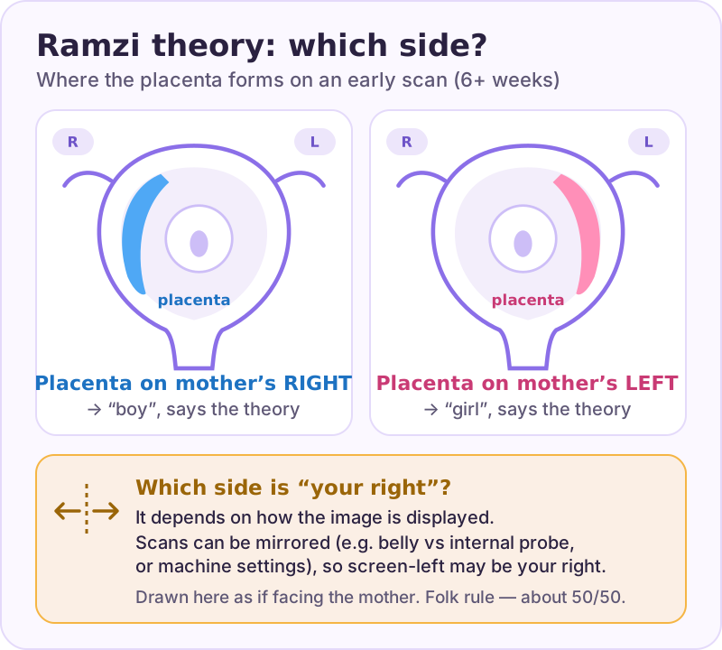
Most 6–8 week scans are transvaginal, with the probe inside. Later scans are usually abdominal, over the belly. The two approaches look at the uterus from different directions. How left and right appear on screen then depends on how the probe is held and how the machine is set up.
Online, you will find rules of thumb such as "transvaginal images are true, abdominal images are mirrored". They are forum conventions, not something established. Settings vary between machines and clinics. A printed picture usually doesn't say which way it is displayed. Unless the sonographer tells you which side is your right, you can't be sure.
That is why Ramzi groups often reach opposite verdicts on the same photo. For more on reading sides, see Ramzi theory: left or right.
Your scan report won't say left or right either
Later scan reports describe the placenta as anterior (front wall), posterior (back wall), fundal (top) or low-lying. These describe front, back and height, not left and right, so they can't be used for Ramzi. The placenta's position relative to the cervix also often looks different later in pregnancy as the uterus grows. That is one reason a low-lying placenta seen early is checked again later.
If you want an answer, here is what works when
| Weeks | Option | How reliable |
|---|---|---|
| 6–8 | Ramzi theory | About chance in the only independent test |
| From about 10 | Prenatal blood test that reports sex (ask your provider) | A medical test, far more reliable than any theory |
| 12–13 | Nub theory | High for specialists on the live scan; unknown from photos |
| 18–22 | Anatomy scan | The standard way to see the baby's sex |
More detail in our week-by-week guide.
How BabyPeek does it
In BabyPeek you measure the nub and mark the placenta side on your own scan, answer the skull-theory questions, and the app adds the Chinese calendar and a playful Photo Pick. It labels how much evidence each method has, shows one majority verdict and turns it into a shareable reveal.
FAQ
Sources
- Ismail SR. The relationship between placental location and fetal gender (Ramzi's method). Contemporary OB/GYN (not peer-reviewed). Source
- P18.17: The role of placental location assessment in the prediction of fetal gender. Ultrasound Obstet Gynecol. 2010 (conference abstract, 277 pregnancies). Source
Written by the BabyPeek editorial team from the studies above. How we research and write. For entertainment, not medical advice.
How BabyPeek does it
BabyPeek reads placenta side as one input among five, then shows a single majority verdict with an honest confidence — so a mirrored image cannot send you the wrong way on its own.