Nub theory, explained
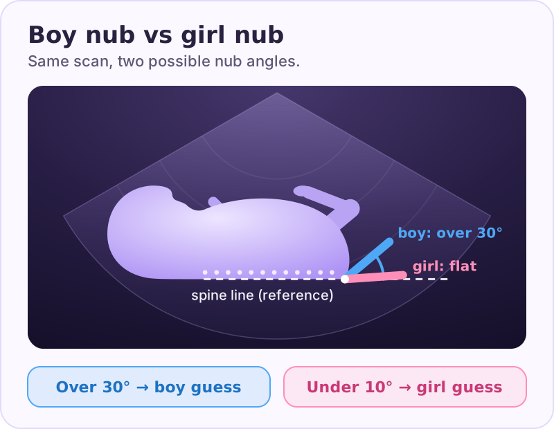
Nub theory predicts your baby's sex from the genital tubercle — a tiny bump near the base of the spine visible on a first-trimester ultrasound. If the nub angles more than 30° away from the spine, it leans boy; if it sits roughly parallel (under 30°), it leans girl. It's a real, peer-reviewed method, but it only becomes reliable from about 12–14 weeks.
What the nub actually is
Early in pregnancy, every baby starts with the same structure: the genital tubercle. It's a small protrusion at the tail end of the spine that will become either a penis or a clitoris. Before about 11 weeks, boys and girls look almost identical down there — which is exactly why early guesses fail.
As the weeks pass, the tubercle of a male fetus tilts upward, away from the spine, while a female's stays flatter, lined up roughly parallel with the body. That difference in tilt is what nub theory reads.
On a good scan you want a clean mid-sagittal (side profile) view — baby's spine along the bottom, the nub clearly separated from the legs. Without that angle, you can't measure anything reliably.
The reason early guesses fail comes down to biology. Until around week 9, the genital tubercle is identical in every fetus; the hormonal signals that differentiate it kick in only gradually. By the late first trimester the male tubercle has begun lifting away from the body, but the female version is still settling into its flatter resting position. That's why the method has a clear "too early" zone and a clear "now it's readable" zone — and why a single week can change your odds dramatically.
How to read the nub: the >30° rule
Want to try it on your own scan? Our free nub angle calculator lets you place the two lines on your photo and measures the angle. The photo never leaves your device.
The core rule comes from the landmark study by Efrat (1999): draw a line through the baby's lower spine, then measure the angle of the nub off that line.
- Angle greater than 30° → likely boy (the "angle of the dangle")
- Angle parallel or under 30° → likely girl
A more recent study put real numbers on the spread: the genital tubercle angle averaged 35.9° in males vs 21.6° in females, and a cutoff of about 28° separated them with 92% sensitivity and 85% specificity (Alfuraih 2024). The boy and girl ranges overlap near the middle, which is why borderline nubs are genuinely hard to call.
Want a deeper walkthrough of the measurement itself? See our guide to the angle of the dangle, and for side-by-side markers read boy nub vs girl nub examples.
Week-by-week accuracy
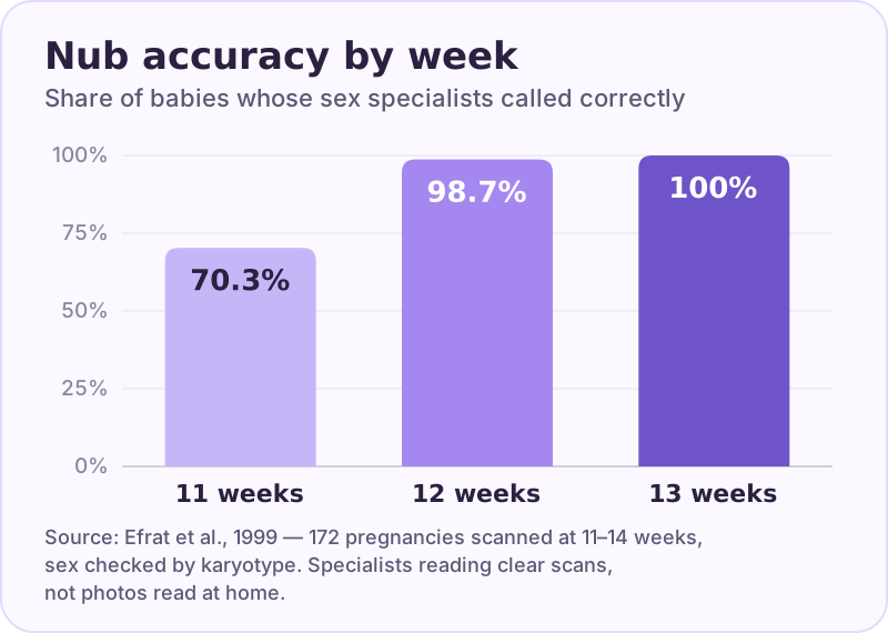
This is the part most people get wrong. Nub theory at 11 weeks is unreliable: overall accuracy was about 70% in the original study. In Efrat's original work, 56% of male fetuses were misassigned as female at 11 weeks — worse than a coin flip — but that error dropped to 3% at 12 weeks and 0% at 13 weeks. Female false positives fell from 5% at 11 weeks to 0% by 12–13 weeks (Efrat 1999).
| Gestational age | Reliability | What's happening |
|---|---|---|
| 11 weeks | Poor (~70%) | Tubercle hasn't tilted enough to tell apart |
| 12 weeks | Good, improving | Male angle becoming clear; some female misreads |
| 13–14 weeks | Best for this method | Angles well separated; cleanest window |
The sweet spot is 12–14 weeks, with 13 weeks often cited as the safest single point. That overlaps neatly with the 11–14-week dating (NT) scan many parents already attend.
Notice an asymmetry in the data, too: boys read more reliably than girls early on. In Efrat's larger follow-up, male predictions were essentially always right by 12 weeks, while female accuracy lagged — 91.5% in the first half of week 12, then climbing to 100% by week 13 (Efrat 2006). So a "boy" call at exactly 12 weeks tends to be sturdier than a "girl" call at the same age, simply because the female tubercle needs a little longer to flatten into its unmistakable position. If your scan suggests a girl right at 12 weeks, a confirming look a week later is reassuring.
Here's the honest version. Under ideal conditions — a skilled reader, a clear mid-sagittal image, and the right gestational age — accuracy is impressive. In Efrat's 2006 follow-up of 656 pregnancies, male predictions were 99–100% correct, and female accuracy climbed from 91.5% in early 12 weeks to 100% by 13 weeks.
But "ideal conditions" is doing a lot of work in that sentence. Real-world results depend on: image quality — a grainy or angled photo can't be measured properly; baby's position — if the legs are crossed or baby is facing down, there's no usable view; and who's reading it — internet strangers eyeballing a blurry photo are not running Efrat's protocol.
It's also worth knowing that researchers have explored a related, sometimes steadier marker: anogenital distance (AGD), the gap measured in that same region. In the 2024 study, AGD averaged about 7.2 mm in males versus 4.4 mm in females and proved a touch more reproducible than the angle itself (Alfuraih 2024). It hasn't replaced the classic angle in everyday "guess the nub" culture, but it's a sign the science is still refining — and that no single first-trimester measurement is a guarantee.
Common mistakes
- Reading a too-early scan. A 10–11-week nub photo tells you very little. Wait for 12 weeks minimum.
- Bad angle. If the view isn't a clean side profile, the "30°" you're measuring may be meaningless.
- Confusing the nub with the cord or legs. The umbilical cord and limbs fool a lot of hopeful parents.
- Treating it as a diagnosis. It's an educated guess, not a medical test. Even a confident nub can be wrong.
- Forgetting the spine reference line — the angle is measured against it, not in isolation.
If nub theory feels too dependent on one tricky angle, you may prefer comparing several signals at once — see how it stacks up against other approaches in our methods compared guide, and read why placental-side methods like Ramzi theory don't hold up under testing.
When a borderline nub is normal
Not every nub falls neatly above or below 30°. The research shows the distributions overlap — male angles average 35.9° and female angles average 21.6°, with real spread on both sides. A nub sitting at 25–32° is genuinely borderline: it's not a failed read, it's just an honest "not sure yet."
A borderline reading most often happens at the edges of the reliable window — right at 12 weeks, or when the scan angle isn't perfectly side-on. In both cases, waiting a week and getting a cleaner image nearly always resolves the question.
See side-by-side examples of clear and borderline cases in our boy-vs-girl examples gallery.
Can an app read the nub?
Reading a nub from a phone photo is exactly where guesses go sideways. The angle is measured against the spine on a clean mid-sagittal image — which is hard to assess by eye, especially from a compressed JPEG photo of a printed scan sheet. Marking the spine line and the nub precisely, then measuring the angle between them, replaces the vague "it looks steep" eyeballing — though the result is still only as good as the image.
BabyPeek lets you mark the nub and spine on your real 12-week scan and measures the angle on YOUR image — not a stranger's eyeballing in a forum thread. It also runs five different methods, including a Chinese-calendar guess and other folk predictors, then gives you a majority verdict so no single tricky angle decides everything.
Best of all, when you're ready, BabyPeek turns the result into a shareable reveal for family and friends. Nub theory is just one signal — the app combines several, and labels how much evidence each one has.
For entertainment only. This is not medical advice — confirm your baby's sex with your doctor's anatomy scan.
How BabyPeek does it
In the app, you mark the nub and spine and BabyPeek measures the angle, then counts it with Chinese, Ramzi, Skull and a playful Photo Pick — and shows one majority verdict with an honest note on each method's evidence.
Frequently asked
Sources
- Efrat Z, Akinfenwa OO, Nicolaides KH. First-trimester determination of fetal gender by ultrasound. Ultrasound Obstet Gynecol. 1999;13(5):305–7. PubMed
- Efrat Z, Perri T, Ramati E, Tugendreich D, Meizner I. Fetal gender assignment by first-trimester ultrasound. Ultrasound Obstet Gynecol. 2006;27(6):619–21. PubMed
- Alfuraih AM et al. The predictive accuracy of anogenital distance and genital tubercle angle for first-trimester fetal sex determination. Diagnostics (Basel). 2024. PubMed
Written by the BabyPeek editorial team from the studies above. How we research and write. For entertainment, not medical advice.